Page 3 - 대한진단검사정도관리협회 뉴스레터 VOL132 (2025년 09월)
P. 3

2025.9_Vol 132  03


                                     정상 또는 전형적 패턴 외에도 다양한 variant signal pattern(예: split signal, atypical fusion 등)이 나
                                     타나며, 이는 clonal heterogeneity를 반영하고 환자의 장기 예후를 설명하는 단서가 될 수 있다.

                                     최근 우리 검사실에서는 기존보다 형질세포 분리, 수율이 향상된 CD138 positive selection kit를 도
                                     입하고, panel에 IGH, MYC break apart probe를 추가하였다. 그 결과, 기존 probe(IGH/FGFR3, IGH/
                                     CCND1, IGH/MAF, IGH/MAFB, CKS1B, D13S319, TP53)에 비해 평균 비정상 검출률이 약 6~8% 증가하
                                     였다. 특히 IGH Dual fusion probe와 IGH break apart probe의 동시 판독은 IGH 유전자 재배열의 다양
                                     한 패턴을 이해하고 결과의 일치성을 확인하는 데 유용하였다.

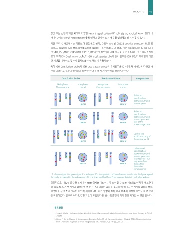
                                     특히 IGH Dual fusion probe와 IGH break apart probe의 동시판독은 IGH유전자 재배열의 다양한 패
                                     턴을 이해하고 결과의 일치성을 보여야 한다. 이에 몇가지 양상을 살펴볼까 한다.

                                             Dual Fusion Probe           Break-apart Probe      Interpretaion

                                         Metaphase      interphase    Metaphase     interphase
                                         Chromosome      nuclei      Chromosome       nuclei


                                                                                               Balanced
                                                                                               translocation
                                                                                               between IGH and
                                                                                               partner gene
                                                         1R1G2F                      1R1G1F

                                                                                               Balanced
                                                                                               translocation
                                                                                               between IGH and
                                                                                               partner gene with
                                                                                               loss of the
                                                         1R0G2F                      1R1G0F
                                                                                               unrearranged IGH

                                                                                               Gain of the
                                                                                               additional copy of
                                                                                               the derivative 14
                                                         1R1G3F                      2R1G1F

                                                                                               Unbalanced
                                                                                               translocation
                                                                                               between IGH and
                                                                                               partner gene due
                                                                                               to deletion of IGH
                                                                                               sequence from
                                                         2R1G1F                      1R0G1F    the partner
                                                                                               derivative
                                                                                               chromosome
                                     * F = fusion signal, G = green signal, R = red signal. (For interpretation of the references to colour in this figure legend,
                                     the reader is referred to the web version of this article.) modified from Chromosomal defects in multiple myeloma.
                                     결론적으로, 다발성 골수종 환자에서 FISH 검사는 여전히 가장 신뢰할 수 있는 세포유전학적 평가 도구이
                                     며, 향후 NGS 기반 검사와 병행하여 통합 진단의 역할이 강화될 것으로 여겨진다. 본 검사실 경험을 통해,
                                     염색체 이상 검출은 단순한 진단적 의미를 넘어 치료 방향과 환자 예후 예측에 결정적 역할을 하고 있음
                                     을 확인하였다. 앞으로 보다 민감한 기술이 도입된다면, 환자 맞춤형 관리에 한층 기여할 수 있을 것이다.


                                        참고 문헌

                                        1.  Sarah E. Clarke , Kathryn A. Fuller , Wendy N. Erber. Chromosomal defects in multiple myeloma. Blood Reviews 64 (2024)
                                         101168
                                        2.  Horna P, Shi M, Olteanu H, Johansson U. Emerging Role of T-cell Receptor Constant   Chain-1 (TRBC1) Expression in the
                                         Flow Cytometric Diagnosis of T-cell Malignancies. Int J Mol Sci. 2021 Feb 12;22(4):1817.
   1   2   3   4   5   6   7